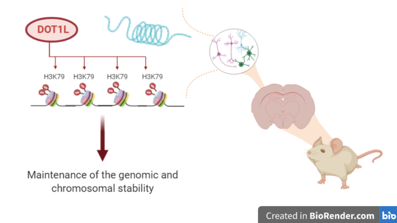
DOT1L affects neuronal layer identity by influencing transcriptional programs
Abstract
A correct cortical development is controlled by epigenetic modifications including chromatin methylation at H3K79 (H3K79me), which is catalyzed by Dot1l-methyltransferase. Dot1l deletion leads to cortical layering defects, indicating H3K79me impact on the cell cycle, progenitors and differentiated neurons.
In eukaryotic cells genetic information is organized into chromatin within the nuclei and its basic units are the nucleosomes. They consist of 146bp DNA wrapped around histone proteins. Histones are subject to post-translational modifications, such as acetylation, methylation, phosphorylation and ubiquitination. These modifications affect chromatin structure by altering the recruitment of DNA binding proteins to particular regions of chromatin, including enzymatic complexes that module transcription. Therefore, heterochromatin is associated with high levels of methylation at H3K9, H3K27, and H3K20, whereas actively transcribed euchromatin is typically enriched with acetylated and methylated H3K4, H3K36, and H3K79 [1].
In a recent work in Nucleic Acids Research Henriette Franz and her collaborators [2] studied the importance of methylated H3K79 in neuronal differentiation. This epigenetic modification is mediated by Disruptor of telomeric silencing-like 1 (DOT1L), a histone methyltransferase capable of catalyzing specifically for H3K79 mono-, di- and trimethylation. DOT1L affects different cellular processes, such as proliferation, DNA repair, and it was shown to have a correlation with the degeneration of some forms of leukemia. However, many underlying molecular mechanisms in the relationship between epigenetic modifiers and central nervous system development were not clear. The authors highlighted the role of DOT1L in progenitors and in differentiated neurons during cortical development.
Franz and collaborators analyzed murine models from the early neurogenesis stage (E12.5) to the adult (E18.5) and through histological images they described differences between the Dot1l-knockout (Dot1l-cKO) line and the wild type line (WT), which was used as a control. Phenotypically Dot1l deletion led to microcephaly and mice died few minutes after birth. Therefore, the scientists tried to discover the underlying molecular mechanism of defects observed during the development of cortical plate in Dot1l-cKO and how Dot1l was related to this phenotype. Using ChIP-seq and RNA-seq, they noticed a significant enrichment of H3K79me2 along the gene body (from the transcriptional start site (TSS) to the transcriptional end site (TES) ± 4 kb) of genes expressed in progenitors (e.g. PAX6, SOX1, SOX2, SOX6, SOX8, SOX10), ventricular zone (VZ), subventricular zone (SVZ) and upper layer (UL) neurons (e.g. SATB2, CUX2, SOX4, SO11), and in just one gene expressed in deep layer (DL) neurons (TBR1). Upon Dot1l-cKO these genes were differentially expressed (DE) and for this reason they showed lower levels of H3K79me2 and decreased expression compared to controls. These data demonstrate that DOT1L targets are characterized by high levels of H3K79me2 upon WT.
Researchers also studied other Dot1l target genes expressed both by progenitors and differentiated neurons, such as HMG-domain Transcription Factors, codified by SOX family genes. In fact, they observed enrichment of H3K79me2 along gene body for some SOX genes and their reduced expression in Dot1l-cKO, by RNA-seq analysis. Therefore, immunostaining experiments revealed a decreased number of progenitors, expressing SOX1, but also differentiated neurons expressing SOX4 and SOX11, while DL neurons expressing SOX5 increased, compared to controls. These results showed that DOT1L regulated HMG-domain TF expression, number of progenitors and differentiated neurons, affecting the cortex layering.
Other analysis involved cell cycle. Researchers, with ChIP-seq and RNA-seq, noted in Dot1l-cKO a decreased expression of peculiar genes of M-phase, marked by H3K79me2 in WT, such as Vangl2 and Cenpv. These are important genes because they regulate the cleavage plan for cellular division. In the early neurogenic phase, when it is important to promote a vertical cleavage plan (symmetric division) in order to maintain progenitor pool [3], they noticed that Dot1l-deficiency in VZ and SVZ decreased expression of M-phase genes, and consequently promoted a premature asymmetric division of progenitors. This led to significant consequences: a modified number of progenitors which implicates an altered number of cortical layers differentiated neurons.

Dot1l deletion leads to a decreased transcription of genes expressed in progenitors and UL neurons at adult stage that is number of cells expressing these genes decreases compared to control, whereas there is no alteration for DL compared to control.
DOT1L deficiency affects in particular the transcriptional programs of both UL neurons and progenitors genes. It means that an alteration, which involves progenitor pool, affects also the other types of cortex cells, and generally, this particular deficiency, starting from the earliest neurogenic phase, reflects to the next ones, resulting in the mutated phenotype.
Considering whole study, Dot1l influences transcription processes, maintaining chromatin accessibility for transcription enzymes. In fact, other studies reported that Dot1l interacts directly RNA Polymerase II on the phosphorylated on C-terminal domain [4], and this could explain the methylation enrichment along the genes bodies, observed in ChIP-seq experiments. Therefore, a set of enhancers dependent on this kind of modification [5] was discovered in acute lymphoblastic leukemia cells expressing the fusion protein MLL-AF4, where the enrichment of H3K79me2 has been found at MLL-AF4-bounding genes: so Dot1l is important to promote enhancer-promoter interactions, contributing again to transcriptional activity. A hypothesis could be that in this compromised clinical situation Dot1l could be a therapeutic target against this disease. Moreover, Dot1l interactions with HMG-domain TF could mean that it promotes the transcription of genes involved in neurogenesis not directly linked with DOT1L as the numerous target genes cited in this study.
This study clarifies the effects of Dot1l deficiency demonstrating how the failure of a single epigenetic factor can provoke serious damages like microcephaly, which is incompatible with life. These effects can be obtained without modifying any DNA sequence but only the chromatin conformation and its spatial organization. However, while the phenotypic effects of Dot1l deficiency are highlighted, there are not specific evidences about molecular mechanisms leading to microcephaly; moreover, we cannot exclude that other epigenetic factors deficiency could provoke cerebral alterations in neuronal development or microcephaly itself, as observed in Dot1l deficiency. So that of course Dot1l is important for transcriptional programs of some cerebral genes involved in the nervous system development, but in general the spatio-temporal activity of epigenetic factors on both chromatin and cell fate is still not totally cleared.
References
- Brendan Jones et al. The Histone H3K79 Methyltransferase Dot1L Is Essential for Mammalian Development and Heterochromatin Structure. PloS Genetics, 2008.
- Franz. et al. Dot1l promotes progenitor proliferation and primes neuronal layer identity in the developing cerebral cortex. Nucleic Acids Research, 2019.
- Anjen Chenn and Susan K. McConnell. Cleavage Orientation and the Asymmetric Inheritance of Notch1 lmmunoreactivity in Mammalian Neurogenesis. Cell, Vol. 82, 631-641, 1995.
- Katherine Wood, Michael Tellier, Shona Murphy. DOT1L and H3K79 Methylation in Transcription and Genomic Stability. Biomolecules, 2018.
- Godfrey et al. DOT1L inhibition reveals a distinct subset of enhancers dependent on H3K79 methylation. Nature Communications, 2018.

